Dacryocystitis
Infection of the lacrimal sac is termed as dacryocystitis. It may result
secondary to the obstruction in the nasolacrimal duct. The lacrimal glands
are located on the upper aspect of the eye in the orbital cavity, each on
both sides towards the lateral portion of the eye.
The lacrimal glands are the specialized exocrine glands responsible for the
production of tears. Tears are the clear fluid that helps in lubricating the
eyes and helps in getting rid of any debris or foreign particles that enter
the eye. Tears are thus the first-line defence for any debris or foreign
particle invasions.
The lacrimal sacs are the reservoirs for the overflow of tears. The
muscular contraction and relaxation of the eyelid muscles helps in the flow
of the tears from the lacrimal sacs. When we blink, our lacrimal sacs pump
the tears inside and outside the eye. This lubricates our eyes and prevents
dryness or itching.
In response to any emotion or any other reflexes, the lacrimal glands
produce tears that flow down and lubricate the eyes, these tears also wash
out the eyes from any debris or bacteria. They then take their way down to
the lacrimal sacs and finally drain into the nasolacrimal duct and get
flushed into the nasal cavity.
You might have noticed whenever we cry more, the tears come from our eyes
and nose at the same time. This is due to the draining process. Similarly,
when we have a runny nose, the eyes are also wet due to the flow of tears.
This is due to the connection between the eyes and the olfactory system.
Nowadays due to the increased screen timing resulting from the overuse of
the mobiles and laptops, eye-dryness has become an annoying problem. Many
ophthalmologists advise us to do regular eye exercises such as eye rotation
techniques, blinking exercises, etc. This is only to activate the lacrimal
glands and lacrimal sacs to pour out tears and lubricate the eyes.
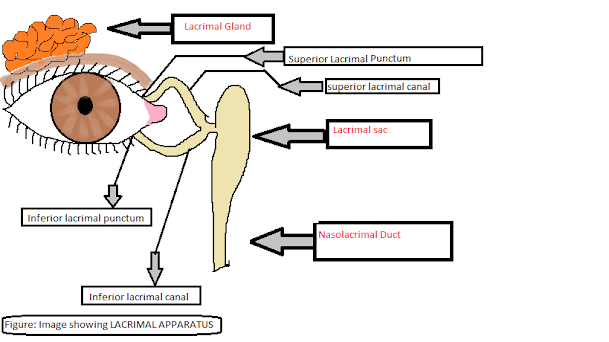
Anatomy of the lacrimal sacs:
Let us take a quick look at the anatomy of the lacrimal sacs and the mechanism of tear production and flow.
Causes of Dacryocystisis
The main causative organisms responsible for the inflammation of the
lacrimal sacs are:
- Staphylococcus aureus
- Streptococcus pneumoniae
- Pseudomonas aeruginosa
The infection is common in infants and people above 40 years of age.
Although it can be found in any one with recurrent flu infections that may
infect the blocked nasolacrimal duct, the acute cases are noted among the
infants. The bacteria cause pus formation in the nasolacrimal duct. It
can be seen outwardly near the medial canthus of the eye slightly near to
the nose. Sometimes the abscess formation is apparent and the doctor may
press it slightly to check whether it contains pus or not.
Based on the onset and the symptoms the condition can be:
- Acute Daryocystitis
- Chronic Dacryocystitis
The other causes or risk factors involved in the inflammation of the lacrimal sacs include:
- Recurrent sinus infections
- Trauma to the nasal bones or adjacent eye structure
- Fractures of the nasal bones leading to blockage of the nasolacrimal duct
- Neoplasms in the nasal or orbital cavity
- Collection of debris inside the sacs
Any of the above factors can possibly infect our lacrimal sacs and cause
its inflammation
The acute cases need medical intervention as the symptoms are more severe
and occur within a short duration following the infection.
The chronic cases are treated with medical as well as surgical
intervention. In case of the recurrent infections, surgery becomes
inevitable to prevent complications and damage to the eyes.
Signs and Symptoms of Dacryocystitis
The person with dacryocystitis presents with following signs and
symptoms:
Pain and swelling near the medial canthus of the eye.
See Also: Conjunctivitis
The present of the purulent swelling apparent on examination
Constant tearing
Redness or erythema over the site of the swelling
Presence of eye discharge in the corner of the eye.
In chronic cases the symptoms are of less severity but they persist for a
longer time and ultimately require surgical approach to get cured.
Diagnosis of Dacryocystitis
The assessment of the signs and symptoms aids in the diagnosis of the
condition. Patient’s previous medical history often helps a lot in ruling
out any possible causes likely to be involved.
An ophthalmologist is an expert in eye diseases and conditions and it is
advisable to consult him/her only when you feel any above symptoms. Some
people may also prefer going to the ENT specialist.
Your doctor may ask you to go for CT scan or culture test for identifying
the causative organism of infection. For culture test, your doctor may press
the swollen area slightly and drain off some pus, and using a swab he takes
the sample to be sent to the medical laboratory for further
investigations.
Treatment Of Dacryocystitis
- Dacryocystitis can be treated with antibiotic eye drops and medications. Drainage of the pus helps in relieving the pressure in the sacs and it may provide some relief.
- After thorough examinations, the doctor may advice to go for medical or surgical intervention.
- The severe cases often need surgery to remove the blockage.
- A surgery called Dacryocystorhinostomy is done to open and drain the blocked duct. In some surgeries, doctors widen the duct passage to allow flow of tears. Sometimes surgery is done to bypass the obstruction and this changes the pathway of the tears away from the blocked portion of the ducts.
Prevention Of Dacryocystitis
The prevention is mostly focused on protecting eye health and avoiding any
possible complications.
- Take appropriate treatment of any flu or sinus-related medical conditions. Consult your physician immediately if you start feeling any bump on the lower side of the eye near to the nose. If you feel your eyes are watering more and it is abnormal according to you, contact your eye specialist and get examined thoroughly.
- Go for surgery in case your doctor has recommended it as the best treatment modality for protecting your eye. Don’t hesitate any ask any queries that brood over in your mind related to the surgery. It is always better to acquire pre-procedure information.
- Eat healthy foods that are rich in Vitamins such as Vitamin A to nourish your eye and enhance its functioning capacity.
- Don’t touch or wipe your eyes constantly.
- Always use a clean cotton cloth or handkerchief to wipe off tears.
- Do not wipe harshly. In this case, as the tears flow excessively you may feel irritated and lose your temper wiping it off every now and then. Wipe out the tears gently.
- Do not touch the swollen area. Always wash your ends with antiseptic soap or liquid before contacting your eyes.


Comments